




-
Single-neuron computation forms the fundamental building block of functional neural circuits. This computation is intrinsically complex, relying on the spatial and temporal integration and nonlinear processing of synaptic inputs across dendritic arbors, followed by thresholding at the axon initial segment1,2. When a neuron fires an action potential, the signal can back-propagate into the dendritic tree, acting as an important feedback mechanism for synaptic plasticity and spike-timing-dependent learning2,3. Visualizing how these electrical signals propagate across the soma and complex dendritic structures is essential for deciphering neuronal computation. However, because dendritic arbors span large three-dimensional volumes, and tracking their dynamics requires high spatiotemporal resolution, such imaging remains technically challenging. While electrical signals themselves propagate too rapidly for large-scale volumetric optical tracking, calcium dynamics provide a widely used indirect proxy. Although calcium indicators exhibit slow decay kinetics4, the timing of their rising phase can be used to map the spatial sequence and directionality of signal propagation. Moreover, calcium imaging captures slower regenerative calcium waves, which play a distinct role in dendritic computation. Nevertheless, accurately resolving the onset of calcium transients throughout extended dendritic arbors remains a demanding task, requiring high-speed volumetric imaging with sufficient spatial resolution to image the dendrite and spine, and sufficient temporal resolution to track rapidly propagating activity.
Most existing approaches of dendritic imaging rely on two-photon calcium imaging5,6 because of its high spatial resolution and optical sectioning capability. However, conventional raster-scanned two-photon microscopy7,8 is fundamentally limited by point-by-point excitation, making it challenging to capture rapid activity propagation across extended dendritic trees. Existing solutions can be broadly categorized into selective-sampling and volumetric projection approaches. Selective-sampling methods improve temporal resolution by interrogating only a subset of the imaging volume. The simplest example is two-photon line scanning9–11, which can achieve kilohertz sampling rates by repeatedly scanning along a predefined dendritic segment, but provides only limited spatial coverage. Acousto-optic deflector (AOD)-based random-access microscopy extends this concept by rapidly targeting multiple user-defined locations, including dendritic branches and spines, enabling simultaneous measurements from distributed compartments12–15. Nevertheless, these approaches are not truly volumetric and generally require predefined regions of interest. In contrast, volumetric projection methods increase volumetric throughput by compressing information from an extended three-dimensional volume into one or a few projections. The most prominent example is Bessel-beam microscopy, where an axially elongated excitation focus simultaneously interrogates structures over an extended depth range16–18. It effectively converts volumetric imaging into a two-dimensional scanning and enables high-speed monitoring of large dendritic arbors. However, signals originating from different depths can overlap at the same lateral location, which introduces ambiguities and signal crosstalk. Related tomographic approaches further use multiple projections and computational reconstruction to recover volumetric information, though at the cost of substantially increased system complexity19. In addition to selective-sampling and volumetric projection approaches, multi-beamlet excitation strategies20–23, in which multiple beamlets with temporal offsets simultaneously scan different regions of the sample, can also increase imaging speed. However, most demonstrations have focused on population-level neuronal imaging rather than imaging dendritic dynamics and signal propagation.
A recent study by Xu et al. introduced dual-view Bessel two-photon projection microscopy (dv-B2PM) for high-speed imaging of dendritic activity24 (Fig. 1). The method extends two-photon Bessel-beam microscopy by simultaneously capturing two orthogonal projections of the same volume, and thereby reduces the structural ambiguities inherent to conventional single-view projection imaging. The underlying optical design is based on a two-photon Bessel-beam microscope. A Bessel beam is rapidly scanned along the fast axis to form a thin two-photon excitation sheet and is then swept through the sample along the slow axis. During this volumetric scan, a conventional projection image is acquired through a photomultiplier tube aligned with the excitation objective. Simultaneously, fluorescence is collected by a camera positioned along an orthogonal detection axis. As the excitation sheet moves through the volume, the camera focal plane is synchronously translated using an electrically tunable lens to track the illuminated plane. Rather than recording individual optical sections which have low signal-to-noise ratios due to short exposure time, the camera integrates fluorescence from successive light-sheet planes during the volumetric sweep, and generates a second projection image from an orthogonal viewing direction. The combination of orthogonal projections enables reliable localization and tracking of calcium activity within sparsely labeled dendritic arbors and allows analysis of dendritic branches that are nearly perpendicular to one projection plane, which would otherwise be difficult to trace using a single-view projection approach. Compared with conventional tomographic systems requiring multiple viewing angles and computational reconstruction, dv-B2PM provides a substantially simpler implementation while retaining three-dimensional localization capability. The system was further integrated with a two-photon glutamate uncaging module for all-optical experiments.
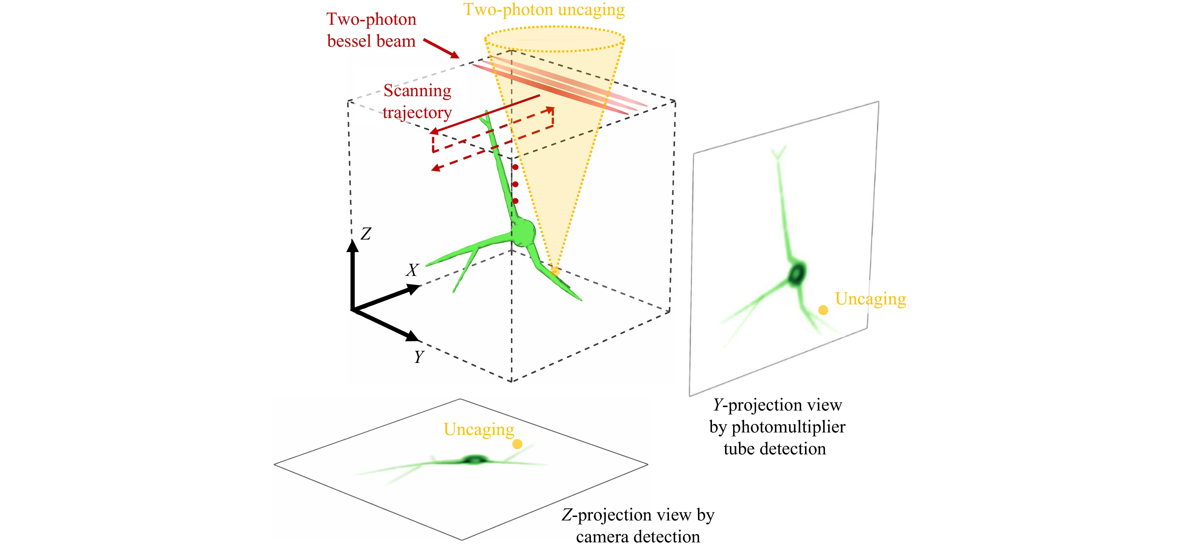
Fig. 1 Schematic of the dual-view Bessel two-photon projection microscope (dv-B2PM) for imaging dendritic calcium activity. A two-photon Bessel beam performs a two-dimensional scan across a three-dimensional tissue volume. Two orthogonal projection views are acquired simultaneously: one by a photomultiplier tube aligned with the excitation path (Y-axis), and the other by a camera—positioned along an orthogonal detection axis (Z-axis)—whose focal plane dynamically tracks the illuminated plane using an electrically tunable lens. A two-photon photostimulation module is integrated into the system for glutamate uncaging to evoke neural activity.
dv-B2PM achieved high spatiotemporal resolution over a substantial imaging volume, enabling volumetric imaging at 100 Hz over 120 × 80 × 42 μm3 with submicron spatial resolution of 0.5 × 0.5 × 0.75 μm3. Using this platform, the authors visualized three-dimensional calcium dynamics evoked by localized two-photon glutamate uncaging in GCaMP8m-expressing mouse brain slices, and tracked calcium signal propagation from the stimulated dendritic region toward the soma. Under suprathreshold stimulation, a delayed calcium component emerged in the soma and subsequently propagated into the dendrites, consistent with calcium signals associated with back-propagating action potentials. Furthermore, spatiotemporal wavelet analysis identified calcium transients spanning multiple frequencies (5−40 Hz), and fast propagating events that reached millimeter-per-second speeds.
While dv-B2PM achieves high spatiotemporal resolution across a relatively large imaging volume, limitations remain. First, the imaging depth is restricted because one of the projection views relies on camera-based fluorescence detection, making image quality increasingly susceptible to scattering and aberrations in deeper tissue. Consequently, the current implementation is best suited for structures located within a relatively shallow depth range along the camera detection axis. Future advances in computational reconstruction and image restoration, particularly algorithms that suppress background fluorescence25,26 and compensate for scattering, may help extend the achievable imaging depth. Second, the dual-view geometry requires optical access from two orthogonal directions, which prevents straightforward translation to in vivo mammalian brain imaging. Future implementations may overcome this challenge by adopting oblique illumination and detection geometries, analogous to swept confocally aligned planar excitation (SCAPE) microscopy27,28, in which both excitation and collection are achieved through a single objective while preserving light-sheet functionality.
The current demonstration of dv-B2PM focuses on visualizing calcium dynamics along individual dendritic branches evoked by localized glutamate uncaging. An exciting future direction will be to combine dv-B2PM with holographic photostimulation29–32 to generate distributed synaptic inputs at multiple dendritic locations simultaneously29,30. Extending the approach from single dendrites to interactions among multiple dendritic branches, and ultimately across neuronal ensembles, could provide a powerful platform for investigating how spatially distributed synaptic inputs are integrated to perform complex dendritic and circuit-level computations.
HTML
-
The author would like to thank Yifei Sun for his assistance in generating part of the figure in this paper.

 DownLoad: