

Special Issue | 22 September 2026
Special Issue on Light and Cancer: Tools for Diagnostics and Theranostics

The goal of this special issue is to present the most exciting developments in the field of light technologies for the early detection, classification and treatment of cancer. This special issue covers the state-of-the-art in this field, as well as the broad and insightful prospects of this urgent technological area, gathering advanced achievements from international research groups and showcasing scientific breakthroughs and practical prospects for the use of light in cancer research and treatment. Special attention will be paid to multimodal approaches that guarantee high accuracy of diagnosis and treatment, where optical technologies can become a powerful addition to high-tech modern imaging modalities such as ultrasound, computed tomography, magnetic resonance imaging and positron emission tomography. Most optical technologies offer great potential for such a combination with the possibility of using fiber optics in clinical settings, including endoscopy.
Currently, optical technologies themselves also provide multimodality in cancer research through the use of various combinations of techniques with different spatial resolution and depth of sensing, which serve as instruments of “optical biopsy”, namely functional near-infrared spectroscopy (fNIRS), diffusion optical tomography (DOT), hyperspectral imaging (HSI), fluorescence lifetime imaging (FLIM), photoacoustic tomography (PAT) and acousto-optic tomography (AOT), multiphoton imaging, optical coherence tomography (OCT), confocal and light sheet microscopies, as well as Raman-based techniques (V. V. Tuchin, J. Popp, and V. P. Zakharov, Eds., *Multimodal Optical Diagnostics of Cancer*, Basel: Springer Nature Switzerland AG, 2020). In turn, approaches to enhancing Raman scattering, such as surface-enhanced Raman scattering (SERS), coherent anti-Stokes Raman scattering (CARS) and stimulated Raman scattering (SRS), can significantly improve the molecular fingerprinting of cancerous tissue for malignancy diagnostics and classification on the molecular level. OCT also offers a diversity of powerful techniques for characterizing cancerous tissues, such as polarization-sensitive OCT (PS-OCT), which allows complementary images to be obtained for the initial polarization and cross-polarization states (CP-OCT), or images for all elements of the complete Mueller matrix or combinations thereof. There are many other OCT tools: label-free OCT angiography (OCTA), OCT-based elastography (OCE), line-field confocal OCT (LC-OCT), Doppler OCT (DOCT), speckle-resolved OCT and others. Current multiphoton imaging systems for in vivo studies use second (SHG) and third (THG) harmonic generation techniques, two-photon microscopy (2PM) and three-photon microscopy (3PM), CARS and FLIM modalities.
This issue is co-edited by
Prof. Valery V. Tuchin, Saratov State University, Russia
Dr. Juergen Popp, Friedrich-Schiller University Jena and Leibniz Institute of Photonic Technology Jena, Germany
Prof. Valery P. Zakharov, Samara National Research University, Samara, Russia
-
Article
Online platform for generating realistic digital phantoms of OCT signals and performing multimodal processing towards optical cancer diagnostics
 Published , Published online: 08 April 2026 , doi: 10.37188/lam.2026.006Optical coherence tomography (OCT), a well-established technique for ophthalmic diagnostics, is now expanding into non-ophthalmic applications, such as dermatology, oncology, and dentistry. OCT signals contain numerous microstructure-sensitive features, including attenuation, speckle statistics, and optical phase. To facilitate the development of feature applications for various tasks and their integration into emerging use cases, we developed a no-code multimodal OCT-integrated online platform for scientific research. This paper describes the capabilities of the developed online platform: realistic digital phantoms of OCT datasets for tuning and benchmarking signal processing approaches; advanced processing of OCT scans to extract feature maps. Several variants of multimodal OCT signal processing have been implemented, including optical attenuation, speckle contrast, depolarisation ratio, strain, and elastographic imaging. This is the first OCT multimodal platform designed to support scientific research aimed at developing various custom-tuned applications, such as disease classification, tumour margin isolation, and severity prediction. We demonstrated the application of this platform for downstream cancer diagnostics using real data from human brain tissue, skin, endometrial tissue, and murine tumour models.
-
Article
Macrophage-mediated delivery of microcapsules for enhanced photodynamic therapy of colon cancer
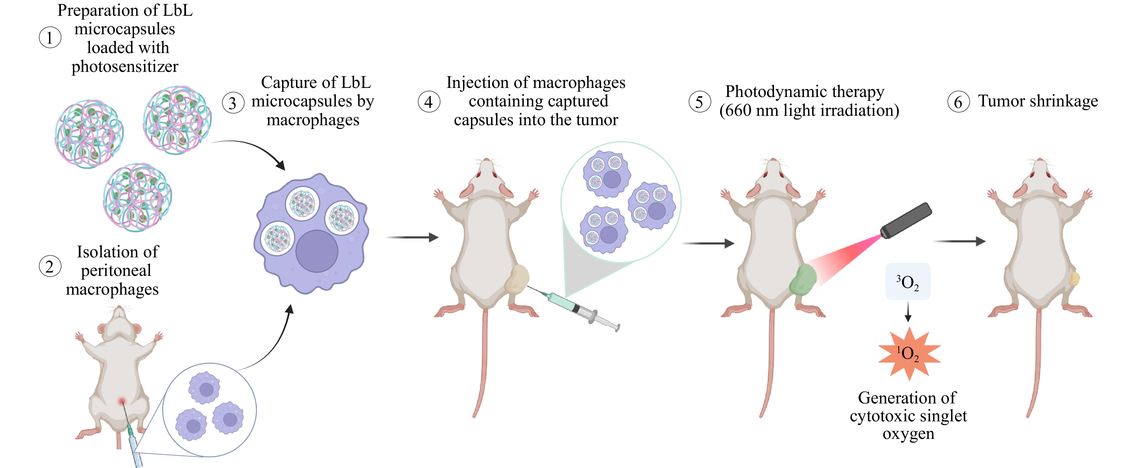 Published , Published online: 12 January 2026 , doi: 10.37188/lam.2026.002Photodynamic therapy (PDT) is a promising strategy for treating solid tumours due to its spatially controlled, light-triggered cytotoxicity. Although recent advances in optical technologies have improved light delivery, PDT efficacy remains limited by insufficient drug accumulation in tumours, largely due to the complexity of the tumour microenvironment. To address this challenge, a macrophage-mediated delivery platform was developed using layer-by-layer (LbL) microcapsules loaded with second-generation photosensitizers: photoditazine (PD) and aluminum tetrasulfophthalocyanine chloride (PS). Both photosensitizers exhibited low dark toxicity and high phototoxicity, enabling their safe transport by carrier cells. The photosensitizers were efficiently encapsulated into LbL microcapsules (6.2 ± 0.5 μm) with different shell compositions. Significant differences were observed between macrophage types: RAW 264.7 macrophages predominantly retained capsules on the cell surface, whereas primary peritoneal macrophages (PMs) internalised capsules within 3 h and retained them for up to 6 d without degradation. Among the tested formulations, polyarginine/dextran sulfate ((PArg/DS)4) capsules loaded with PD demonstrated the highest uptake efficiency and supported macrophage migration into tumour spheroids. In vivo experiments using a CT-26 colon cancer model confirmed the therapeutic potential of this platform, while highlighting the need for further optimisation for large tumours. This study provides new insights into cell-mediated delivery systems and underscores their potential to enhance PDT outcomes beyond current limitations.
-
Article
Air-coupled ultrasound based noncontact reverberant optical coherence elastography
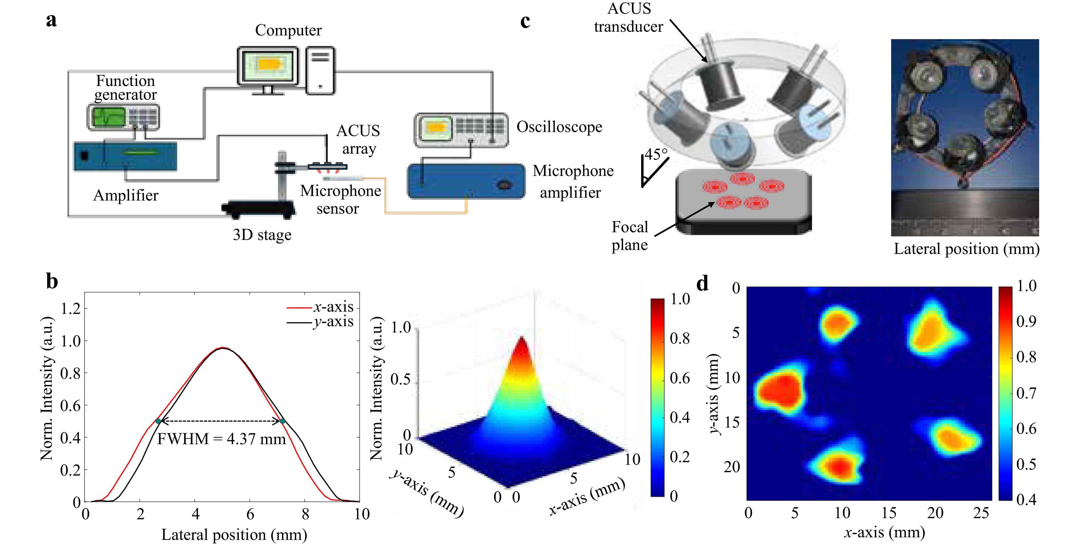 Published , Published online: 09 December 2025 , doi: 10.37188/lam.2025.076Reverberant optical coherence elastography (Rev-OCE) has been used to map the mechanical properties of tissues with high quality and resolution regardless of tissue boundaries and stiffness. Generally, Rev-OCE utilizes the interference of multiple arbitrary mechanical waves generated by distinct sources in direct contact with the sample. In this study, a novel methodology was designed and implemented to create a completely noncontact method utilizing multiple air-coupled ultrasound (ACUS) transducers capable of generating a reverberant field within the sample. An array of unfocused ACUS transducers with a resonant frequency of 40 kHz was characterized and placed in a 3D-printed ring to produce a reverberant field in a side-by-side gelatin phantom (4% and 8% w/w gelatin concentration). The results showed the generation of a reverberant field within the sample with a transverse resolution of 208.8 ± 72.4 μm. Also, this method was tested in porcine cornea showing the stiffness differences found in the corneal layers, where the wave speed is substantially higher in the epithelium (3.7 m/s) and anterior stroma regions compared with the posterior and endothelium regions (2.2 m/s). The generation of a reverberant field using ACUS Rev-OCE guarantees the integrity of sensitive samples, and it shows its potential to be used in-vivo without causing any damage or discomfort in live subjects.
-
Article
QPI- and FLIM-assisted multimodal analysis of the dynamics of morphological and physiological parameters of cells
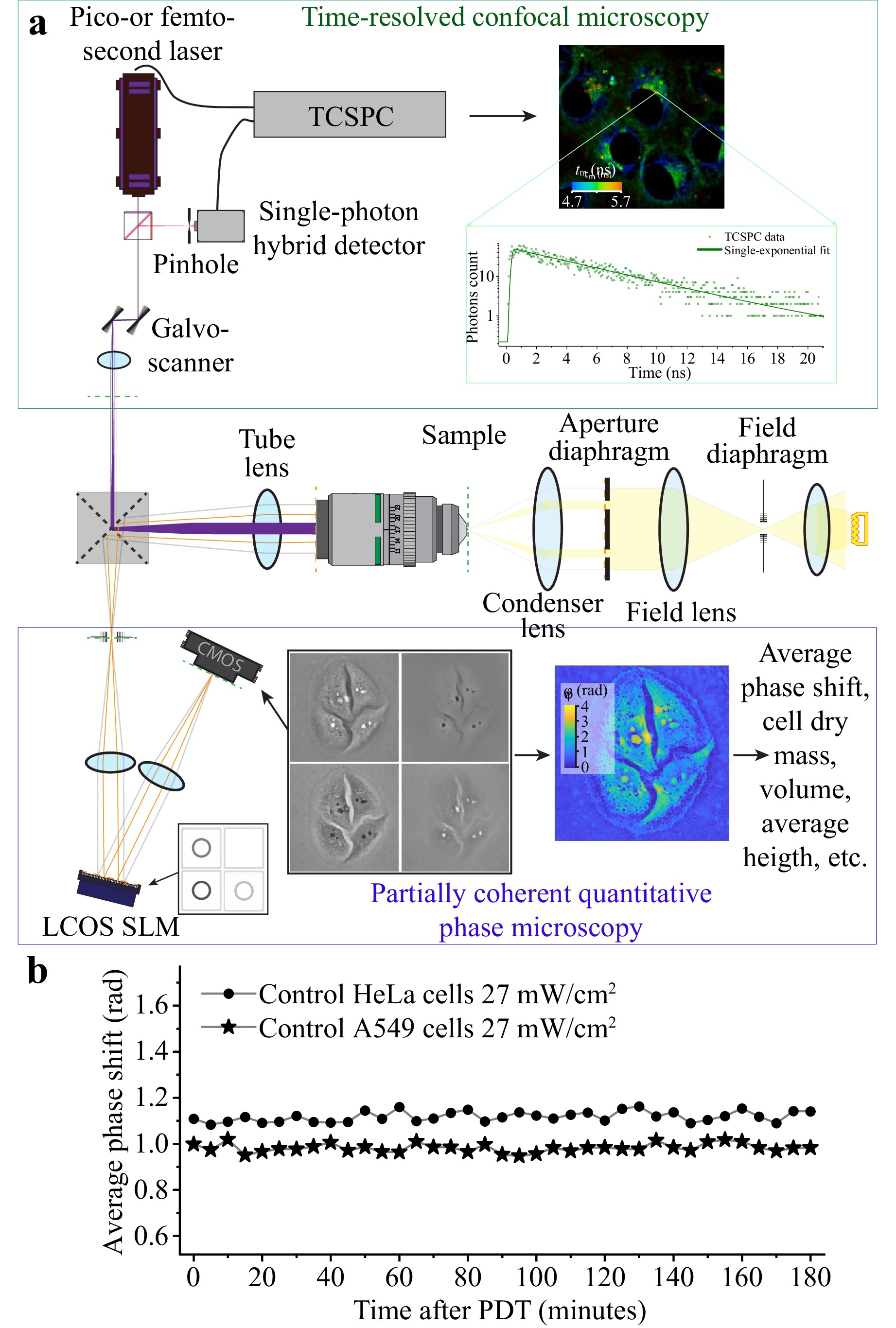 Published , Published online: 25 November 2025 , doi: 10.37188/lam.2025.079This paper presents a multimodal approach that combines time-resolved fluorescence microscopy and spatial light interference microscopy into a single complex allowing for a concurrent comprehensive analysis of cell samples in vitro. The combined application of quantitative phase imaging using partially coherent radiation with time-resolved fluorescence imaging of endogenic coenzymes NADH and FAD, as well as Radachlorin photosensitizer, allowed for evaluation of a set of morphological and physiological parameters of cells of the two lines, HeLa and A549, in the course of photodynamic treatment. Besides comparison of cells of the two lines in terms of volume, height, dry mass, projected area, and redox ratio, we analysed the mechanisms and rate of Radachlorin accumulation. Using the developed multimodal approach, we demonstrated that in both the cell lines, Radachlorin was taken up in complexes with serum albumin, transported by endocytosis, and accumulated primarily in lysosomes. The Radachlorin transportation through the cellular membrane to lysosomes was accompanied by a decrease in its fluorescence lifetime, which was apparently owing to a gradual increase in endosome acidity. The different contributions of the fast and slow components of time-resolved fluorescence signals of nicotinamide adenine dinucleotide (NADH) and flavin adenine dinucleotide (FAD) enabled us to demonstrate the difference in the redox ratios of cells of the two lines. The average concentration of the accumulated Radachlorin photosensitizer in cells of the two lines and their resistivity to photodynamic treatment also differed: HeLa cells tended to accumulate more Radachlorin and demonstrated lower resistivity to photodynamic treatment.
-
Article
Bimodal tissue differentiation using hyperspectral imaging and elastographic Fourier transform profilometry
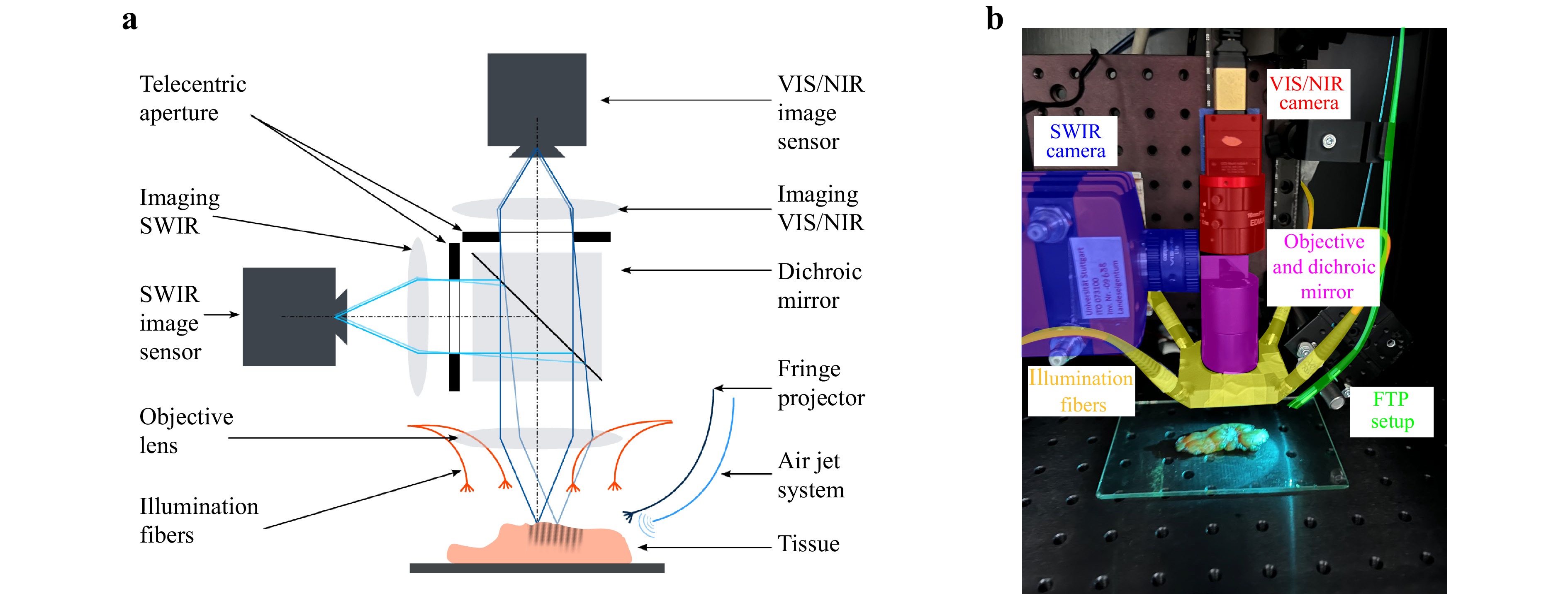 Published , Published online: 06 November 2025 , doi: 10.37188/lam.2025.073Multispectral imaging is a common method to enhance tissue differentiation based on different cell or tissue structures. These differences arise from the unique structural properties of each tissue type, which result in characteristic differences in spectral absorption. In the context of distinguishing between healthy and malignant tissues, differences are observed not only in spectral absorption but also in their elastic properties. To improve the reliability of measurement devices, we present a novel bimodal measurement system that includes a hyperspectral and an elastographic Fourier transform profilometry system to combine the individual results and to improve the overall task of tissue differentiation. The results of both subsystems show good ability to differentiate between healthy and malignant tissue. However, some limitations were identified for each individual sensor concept, such as contamination with pathological dye in hyperspectral imaging and insufficient sample thickness in elastographic measurements. Nevertheless, the multimodal combination of the individual sensor systems ensures good tissue differentiation. While the elastographic sensor can differentiate tissue even when contaminated with pathological staining agents, hyperspectral imaging can be used on very thin tissue samples and also enables the detection of tumor boundaries.
-
Article
Optical percutaneous needle biopsy in oncology
 Published , Published online: 31 October 2025 , doi: 10.37188/lam.2025.072New technologies that combine standard procedures for tissue sample extraction and analysis with optical spectroscopy and imaging represent a new step in the development of tumour tissue diagnostics. Optical percutaneous needle biopsy is a promising method for increasing the diagnostic efficiency of the procedure by selecting the area for sample extraction and obtaining information about vascular changes and the metabolism of biological tissues at the needle tip. This study provides a comprehensive analysis of existing technical solutions to address various diagnostic tasks: studying the metabolic status of tissues for the adjustment of treatment protocols, differentiating healthy and tumour tissues for navigating puncture needles, and obtaining preliminary diagnoses of the nature of neoplasms. Technical systems for assessing the perfusion and metabolic characteristics of biological tissues using fine-needle probes have been described. Fine-needle probe designs for biopsies of various organs are also presented. The results of using optical percutaneous needle biopsy in liver and breast cancers are demonstrated.
-
Review
Terahertz endoscopy of hard-to-access objects in the context of neoplasms diagnosis–A review
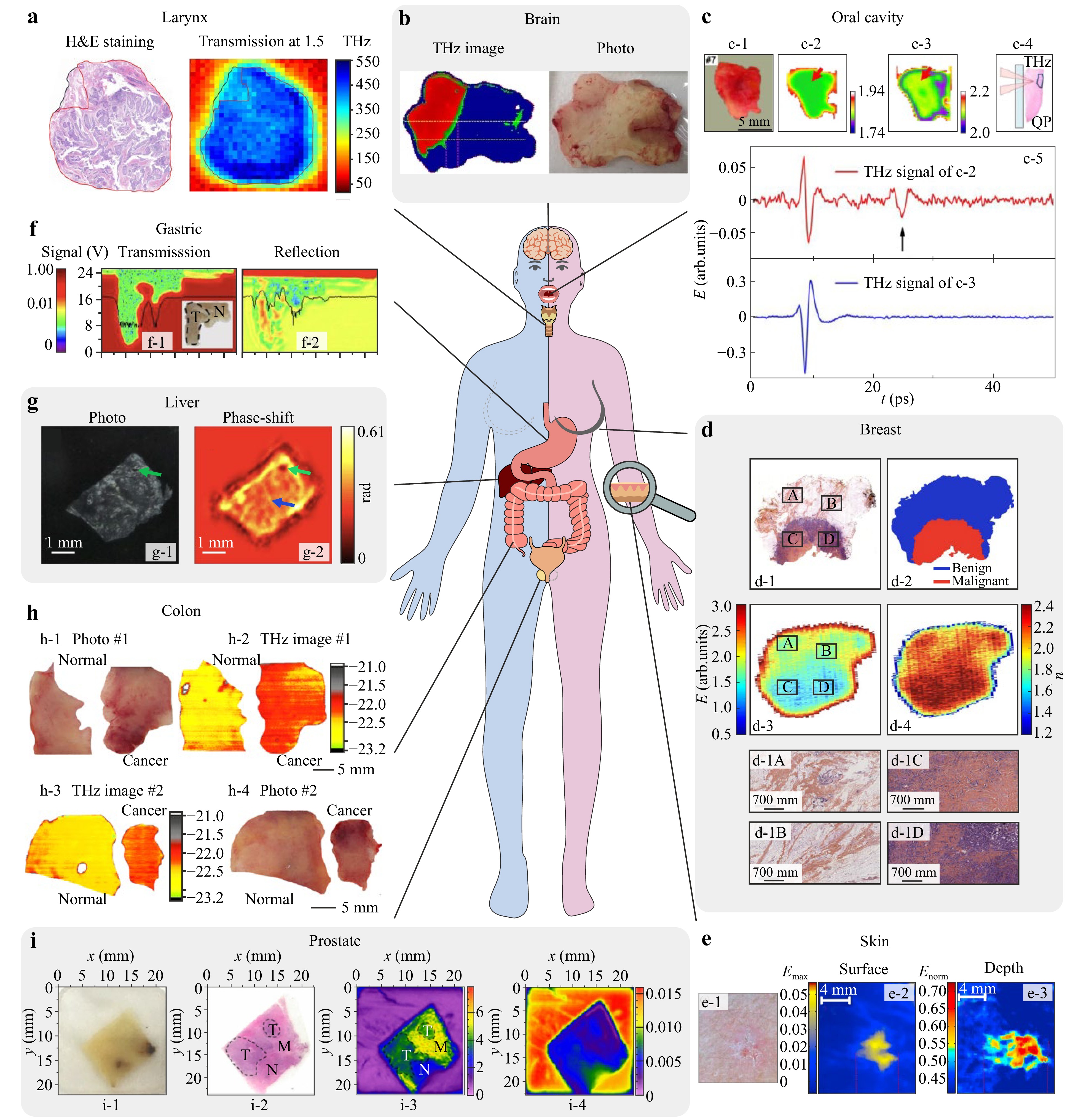 Published , Published online: 21 August 2025 , doi: 10.37188/lam.2025.058Although terahertz (THz) spectroscopy and imaging offer a variety of applications in medical diagnosis of malignant and benign neoplasms, their translation into clinical practice is hampered by the absence of endoscopic systems capable of sensing the THz optical properties of the hard-to-access tissues. In this review, we focus on recent attempts to address this challenge. To better highlight the need for THz endoscopes, we start with a brief overview of THz medical applications, with an emphasis on neoplasms diagnosis. We then consider the two existing principles of THz endoscopy. The first uses the fiber-coupled THz photoconductive antennas (PCAs) for the THz generation and detection in close proximity to a hard-to-access object, where optical fibers are applied to flexibly deliver the laser pump and probe beams to the THz emitter and detector. The key technology of the second approach is the THz optical fibers capable of delivering the THz waves to an analyte and then detecting the reflected and back-propagated THz signal. Despite this approach still lacking the efficient commercially available THz fiber optics, most recent developments pave the way to solve these problems. In this review, several notable examples of THz endoscopic systems based on different guiding mechanisms, material platform, and manufacturing strategies are discussed.
-
Review
Application of fluorescence lifetime imaging-integrated deep learning analysis for cancer research
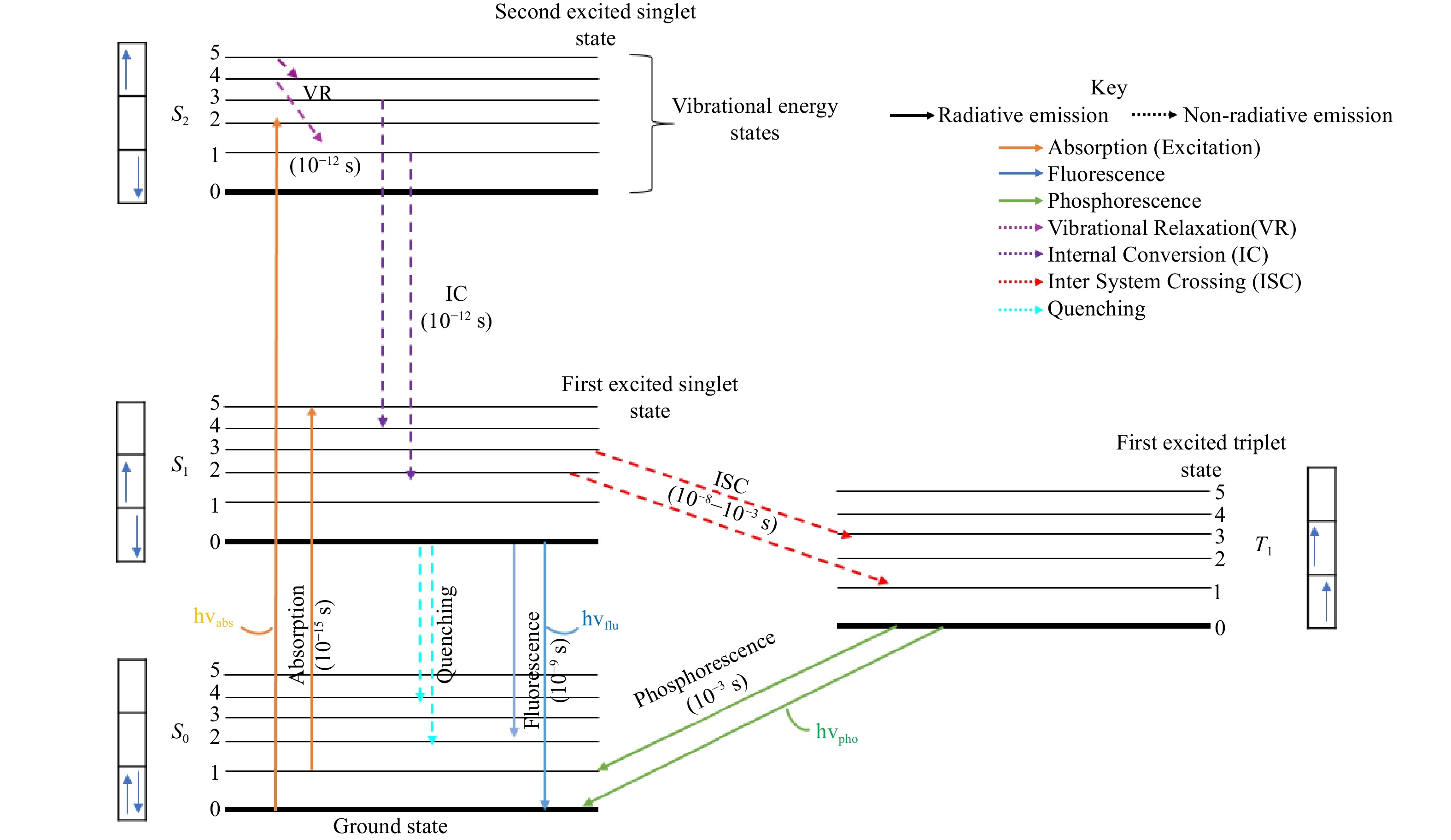 Published , Published online: 05 August 2025 , doi: 10.37188/lam.2025.049Fluorescence lifetime imaging microscopy (FLIM) has emerged as a transformative imaging technique in cancer research, offering quantitative insights into cellular metabolism, tumor microenvironments, and therapeutic responses. By measuring the fluorescence lifetimes of metabolic cofactors such as NADH and FAD, FLIM facilitates the analysis of cancer-specific metabolic reprogramming and heterogeneity. Integration with deep learning further enhances FLIM’s diagnostic and therapeutic potential, enabling high-resolution imaging, automated data analysis, and biomarker identification. This review provides a comprehensive overview of the principles and technological advancements of FLIM, highlighting its applications in cancer diagnostics, drug delivery, and therapy, as well as its integration with deep learning to increase imaging precision and data interpretation. Challenges such as high costs, high computational complexity, and the need for standardized imaging protocols are also addressed. By bridging FLIM with cutting-edge computational techniques, this review highlights its potential to revolutionize cancer research, paving the way for early diagnosis, personalized therapies, and deeper insights into tumor biology.
-
Article
SERS-based technique for accessible and rapid diagnosis of multiple myeloma in blood serum analysis
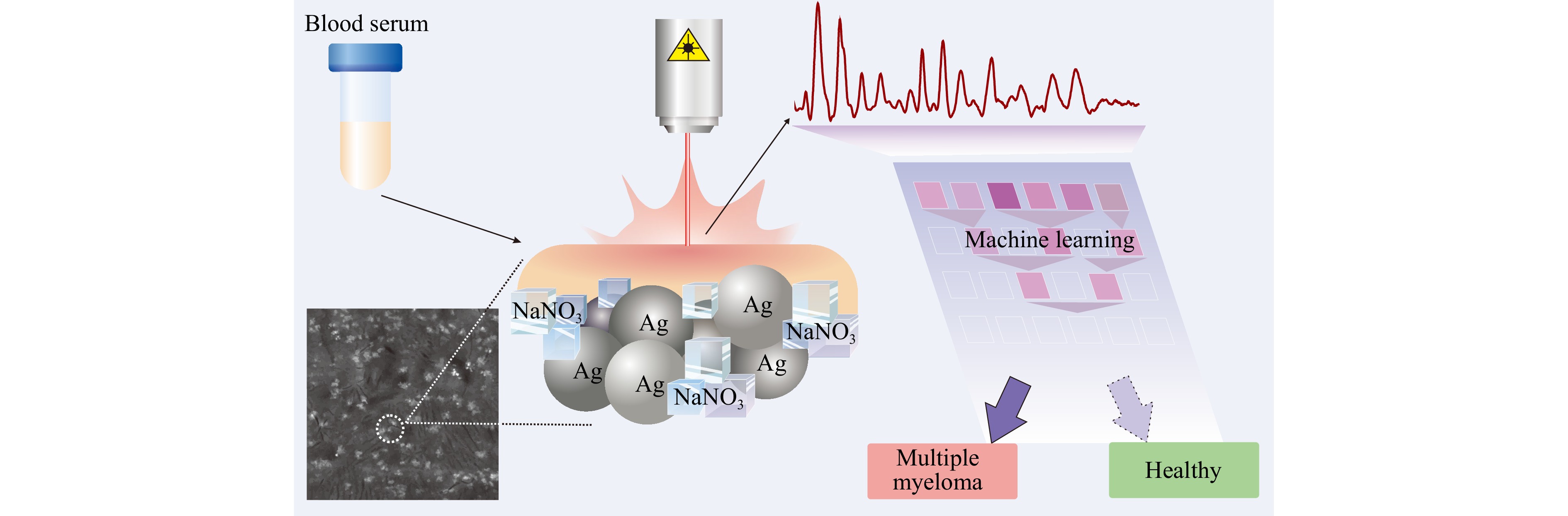 Published , Published online: 13 May 2025 , doi: 10.37188/lam.2025.035Surface-enhanced Raman spectroscopy (SERS) is a promising analytical method for studying the structure and composition of multi-component media in clinical practice. However, the practical application of SERS imposes several conditions and restrictions depending on the required analytical accuracy and implementation complexity. This paper proposes a simple serum SERS technique for diagnosing multiple myeloma (MM). The process utilises a substrate composed of agglomerated spherical silver particles (~200 nm) with a capillary effect on sodium nitrate crystals (0.02% volume concentration), excited at a wavelength of 785 nm. Spectral characteristics were recorded using a detector without external cooling, ensuring a cost-effective approach. The proposed low-cost approach was tested on 31 samples from MM and 102 serum samples from an age-matched control group. Combining the SERS technique with the multivariate analysis for serum testing provided high discrimination rates for MM patients compared to the control patients, with an average accuracy of over 96%. Biochemical interpretation of the recorded spectra identified the informative bands at 635 cm-1, 723 cm-1, and 1052 cm-1. Overall, the proposed SERS-based tool can become the basis for screening for MM and can be easily integrated into clinical practice, expanding diagnostic capabilities where expensive laboratory techniques are not affordable.
-
Article
A comparative study of plasmonic nanoparticles for targeted photothermal therapy of melanoma tumors using various irradiation modes
 Published , Published online: 11 March 2025 , doi: 10.37188/lam.2025.005
Melanoma, a highly malignant and complex form of cancer, has increased in global incidence, with a growing number of new cases annually. Active targeting strategies, such as leveraging the α-melanocyte-stimulating hormone (αMSH) and its interaction with the melanocortin 1 receptor (MC1R) overexpressed in melanoma cells, enhance the concentration of therapeutic agents at tumor sites. For instance, targeted delivery of plasmonic light-sensitive agents and precise hyperthermia management provide an effective, minimally invasive treatment for tumors. In this work, we present a comparative study on targeted photothermal therapy (PTT) using plasmonic gold nanorods (Au NRs) as a robust and safe nanotool to reveal how key treatment parameters affect therapy outcomes. Using an animal model (B16-F10) of melanoma tumors, we compare the targeting abilities of Au NRs modified with two different MC1R agonists, either closely mimicking the αMSH sequence or providing a superior functionalization extent of Au NRs (4.5% (w/w) versus 1.8% (w/w)), revealing 1.6 times better intratumoral localization. Following theoretical and experimental assessments of the heating capabilities of the developed Au NRs under laser irradiation in either the femtosecond (FS)- or nanosecond (NS)- pulsed regime, we perform targeted PTT employing two types of peptide-modified Au NRs and compare therapeutic outcomes revealing the most appropriate PTT conditions. Our investigation reveals greater heat release from Au NRs under irradiation with FS laser, due to the relaxation rates of the electron and phonon temperatures dissipating in the surrounding, which correlates with a more pronounced 17.6 times inhibition of tumor growth when using FS-pulsed regime.
Prof. Valery V. Tuchin
Saratov State University, Russia
Valery V. Tuchin is the corresponding member of the Russian Academy of Sciences, Head of the Department of Optics and Biophotonics and Director of the Science Medical Center of Saratov State University. He is also works with several other research centers of RAS and universities. His research interests include biophotonics, tissue optics, drug delivery, and phototherapy. He is the Honored Scientist of Russia, Fellow of SPIE and OPTICA, Distinguished Professor of Finland, recipient of SPIE Educator Award, Joseph W. Goodman Book Writing Award (OPTICA/SPIE), Michael S. Feld Biophotonics Award (OPTICA), Chime Bell Award of the Chinese province of Hubei, the D.S. Rozhdestvensky Medal and the S.I. Vavilov Medal of the Russian Optical Society, and the A.M. Prokhorov Medal of the Russian Academy of Engineering Sciences.

Dr. Juergen Popp
Friedrich-Schiller University Jena and Leibniz Institute of Photonic Technology Jena, Germany
Since 2002 Juergen Popp is holds a chair for Physical Chemistry at the Friedrich-Schiller University Jena, Germany. Since 2006 he is also the scientific director of the Leibniz Institute of Photonic Technology, Jena. Juergen Popp is a world leading expert in Biophotonic / optical health technology research covering the complete range from photonic basic research towards translation into clinically applicable methods. He has published more than 990 journal papers, has been named as an inventor on 15 patents and has given more than 200 invited talks on national and international conferences (among them more than 50 keynote/plenary lectures). He received several awards like e.g. the 2016 Pittsburgh Spectroscopy Award, the Kaiser-Friedrich-Forschungspreis 2018, the third price of the Berthold Leibinger Innovationspreis in 2019 or in 2022 he became part of The Photonics100. Furthermore, he received two honorary doctorate degree: 2012 from the Babes-Bolyai University in Cluj-Napoca, Romania and very recently 2023 from the University at Albany – State University of New York (USA).

Prof. Valery P. Zakharov
Samara National Research University, Samara, Russia
Valery P. Zakharov is a Professor and Chair of Laser and Biotechnical Systems Department, Head of Photonics Laboratory at Samara National Research University. His research interests include laser medicine, biophotonics, multimodal biomedical imaging, and application of fluorescence and Raman-techniques for biomedical diagnostics. He has published more than 350 publications in peer-reviewed journals, has several patents and yearly has given invited talks on national and international conferences. He has been awarded Honorary Worker of Education of Russian Federation, Samara Award for Biophotonics innovations.

 Email
 RSS
